The difficulty for a breeder or a trainer is to obtain a good quality of bones and other major structures of the locomotor system such as tendons, muscles, and ligaments in his horses, through an adapted training and a balanced diet. It is also important to remember that some horses start out deficient because of poor conformation that induces abnormally high forces on certain bones and joints, making them more susceptible to developing ailments.
Structure, function, and adaptation of the horse’s bone
Bones can be classified by size, shape, location, or function. One especially important type of bone in a horse’s skeleton is the long bone. This type of bone has a complex architecture and organization. It is surrounded by a periosteum, a fibrous membrane richly vascularized and containing osteoblasts capable of repairing lesions on the bone surface. This surface is made up of a very dense compact bone that envelops the spongy or trabecular bone, which is made up of bone trabeculae that protect the bone marrow. This marrow can take two forms: the yellow or fatty marrow and the red marrow which is the source of red and white blood cells. The bone tissue, which is compact or spongy, is continually active and is subject to perpetual remodeling throughout the horse’s life. This tissue is made up of a matrix containing cells (osteoblasts, osteocytes, osteoclasts) on which a mineral phase based on calcium and phosphorus is deposited.
Bone, which is thus physiologically reworked in a perpetual way, is also capable of responding to mechanical stimuli thanks to a complex organization between the several types of bone cells. Overall, exercise leads to an increase in bone density by increasing the deposition of the mineral phase, whereas, conversely, inactivity or immobilization of a limb by a cast will reduce bone density.
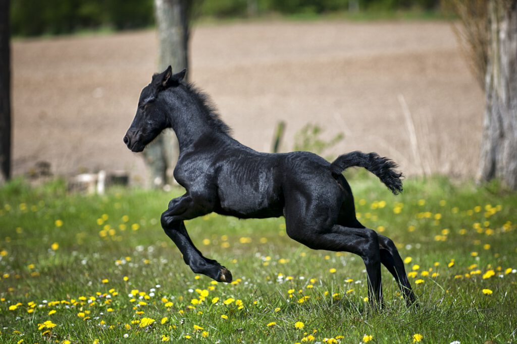
In the horse, the forces induced by exercise are exerted on the bones of the four limbs. It is here that many skeletal adaptations are observed.
In some cases, the response of the bone tissue may be excessive in either direction, resulting in pathological changes to the bone ranging from osteophytes or exostoses (new production of bone tissue on the surface of a normal bone), sclerosis (excessive increase in density), demineralization (loss of bone density), to partial or complete fracture of a bone. A typical example of exostosis is the “suros” regularly observed in the region of the cannon. Demineralization will be observed when a limb is not used or when a bone is chronically compressed.
Fractures in horses
Fractures in horses can affect any bone. Some are related to an adaptation problem (fatigue fractures) and others are caused by direct trauma to the bone. It is essential to define whether a fracture has joint involvement or not and whether there is soft tissue breakdown on the surface of the fractured bone. If there is, it is called an open fracture; it is always an emergency and the prognosis is, regardless of the location of the fracture, much worse than for a closed fracture of the same site. Fracture-related symptoms vary depending on the location, type, and extent of the fracture.
Complete fractures of a bone are mostly accompanied by displacement and are easy to recognize because of the sharp pain on palpation, the swelling that will quickly develop around the fracture site, the lack of support of the limb on the ground or the evidence of crepitations. Other types of fractures are sometimes more difficult to recognize. For example, an incomplete fracture (e.g., a fatigue fracture), often observed in galloping horses, will induce signs that are less marked but amplified with exercise. A horse with an intra-articular fracture will have a variable symptomatology depending on the type of fracture. In the case of a small fragment, commonly called a joint mouse, distension of the joint with moderate lameness will be observed, whereas a so-called in bloc or comminuted fracture, which involves a larger part of the joint surface, will cause much more significant lameness. In all cases of fracture, the complementary examination of choice is radiology. This examination is essential to objectify the extent of the lesions and to prepare for surgery if it is necessary.
How to treat a fracture in the horse
The treatment of a fracture must consider a basic principle which is to obtain a good stability of the fracture site to allow an excellent quality bone repair. If this is achieved, the pain will be reduced immediately and after about two months, fusion between the bone ends will be achieved. The return to normal strength will take between six and twelve months. A combination of rest, immobilization of the involved limb with a cast or support bandage, and appropriate surgery is most often used to achieve stability. Surgery consists of either arthrotomy or arthroscopic excision of the fractured fragment if it is small or the use of internal or external fixators in other cases. Internal fixators such as screws and plates are most often used in horses. Once the fracture is repaired, it is particularly important to resume controlled and very progressive exercise so that the bone can adapt to the type of work required.
The vital prognosis in adult horses, or those weighing more than 300 kg, is currently poor for many fractures involving load-bearing skeletal structures such as the long bones of the limbs. However, the prognosis is often good for smaller fractures or fractures on bones that are not used for support (rudimentary metacarpals, ulna), and the prognosis is variable for bones under stress (navicular bone, patella, sesamoids) because they heal poorly and slowly. However, overall, the prognosis for life and sport is constantly being improved thanks to the constant progress in surgical techniques and implants used in equine medicine.
Since the 1990s, veterinarians have also been working to prevent bone problems rather than cure them. This is done through the search for non-invasive techniques to evaluate the skeleton. For example, molecules that can be measured in the blood are markers of bone turnover, and ultrasound shows promise in assessing bone structure and density. The work of an equine osteopath can also prevent and cure certain bone diseases in horses.
Osteomyelitis and sequestration in horses
Osteomyelitis is a bacterial infection of the bone with destruction of the bone and very often anarchic bone proliferation. The horse with this type of problem will present with moderate lameness, the infected area will be warm, painful and a purulent discharge will be present if the infection is secondary to trauma. A swab of the discharge or a bone biopsy of the infected area will be submitted for bacteriological examination to isolate the germ involved. An X-ray and/or ultrasound examination is recommended to assess the extent of the damage and to determine whether there is a bony sequestrum, i.e., a devitalized bone fragment. It should be noted that a bone sequestrum can develop without infection and be the result of a local absence of vascularization of the bone, as in the case of deep lacerations with removal of the periosteum of the underlying bone. It will take two to three weeks to properly observe a bone sequestrum on radiological examination.
The principle of treatment of a bone infection and/or sequestration, whether septic or not, is a wide resection of all abnormal bone tissue. General antibiotic therapy is combined with local or regional application of antibiotics. In case of severe lameness, support of the opposite limb with bandages should also be considered. In foals, osteomyelitis is very often related to septic arthritis following sepsis, which in most cases originates in an umbilical abscess. In this case, in bloc resection of the umbilicus and related structures is mandatory for successful treatment of the osteomyelitis.
If the infection affects a superficial bone, the prognosis after treatment is usually particularly good for sport, whereas if the infection is present at a fracture site or following sepsis in the foal, the prognosis is much more reserved, not only for sport but also for life. In the foal, irreversible bone deformities may remain.
Other bone diseases in horses
Metabolic diseases or tumors of the bone in horses are rare and only two examples will be presented here: juvenile mandibular ossifying fibroma and aneurysmal cyst of the bone, so that the reader is aware that a multitude of bone diseases have also been described anecdotally in this species. Juvenile mandibular ossifying fibroma is a benign proliferative and exuberant tumor of the bone involving the rostral region of the mandible of young horses. It is disfiguring and is often accompanied by significant ulceration of the hyperplastic gingiva. The recurrence rate is high if the mass is not completely excised. The treatment of choice is therefore a single or bilateral mandibulectomy (removal of the rostral part of the lower jaw). The prognosis is then favorable. The aneurysmal bone cyst is a benign lesion, usually localized but invasive. Its pathogenesis, congenital or secondary to trauma or a neoplastic process, is not established; the development of the lesion is often associated with hemodynamic abnormalities such as venous obstructions or arteriovenous fistulas. Treatment is surgical excision.