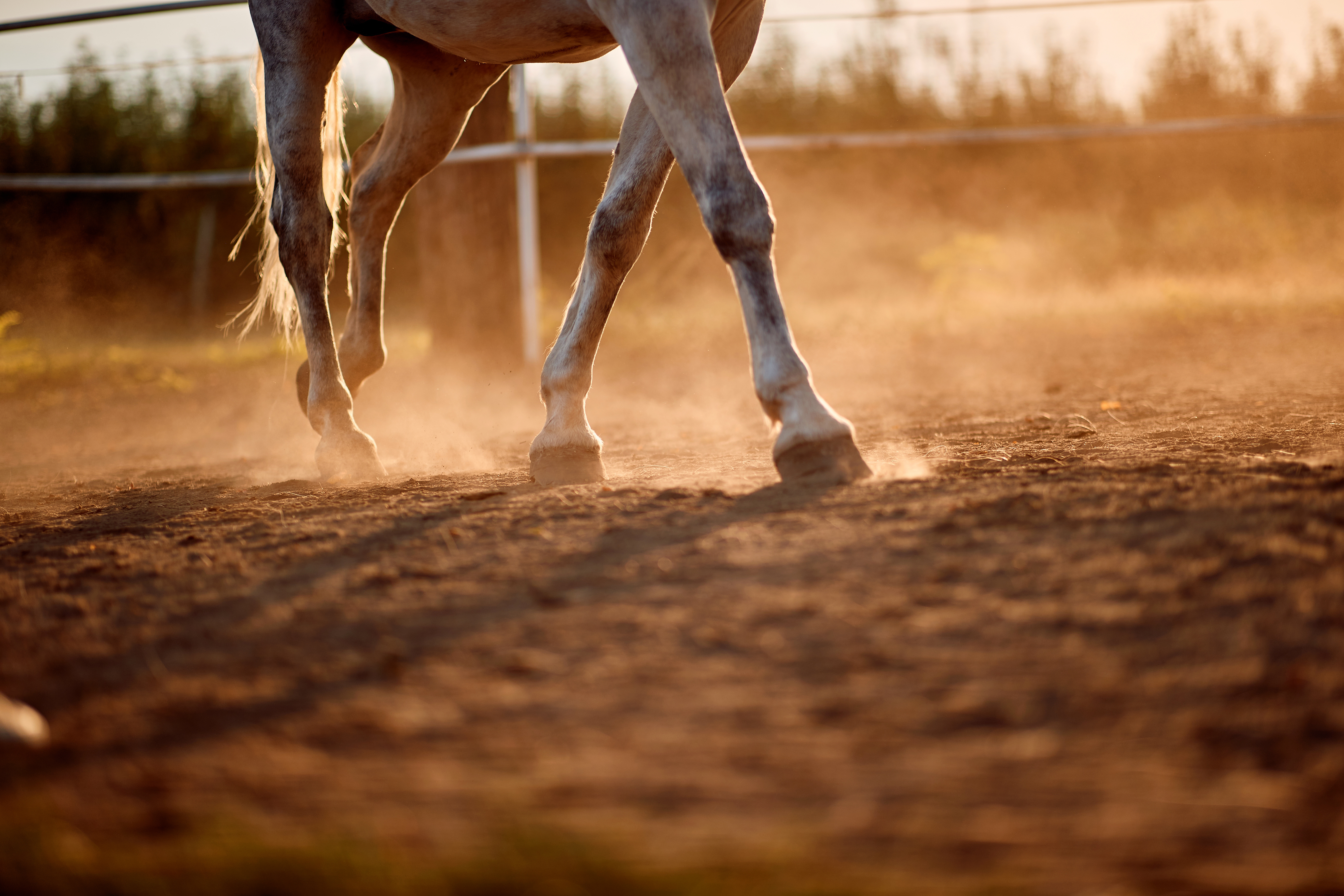
Laminitis is a condition that involves one or more of a horse’s feet. The foot consists of a horny box, rigid on its wall, more flexible and closed in its distal part (sole and frog). It contains the bone of the 3rd phalanx, the navicular bone, the foot joint, the complementary fibrocartilage of the third phalanx and all the peripheral soft tissues. The link between the corneal box and the soft tissues is made by imbrication between the lamellae of the podophylla (sensitive tissue located on the surface of the 3rd phalanx) and the lamellae of the keraphyll (deep layer of the corneal box which is insensitive). The foot is delimited in its upper part by the coronary band characterized by the presence of a strong vascularization and cells responsible to produce the hoof horn.
Laminitis in horses
Laminitis is a condition that involves one or more of a horse’s feet. It occurs in two main forms: with a rotation of the 3rd phalanx or with a descent of the phalanx. The etio-pathogenesis is still poorly understood, but the result is significant inflammation, with separation between the foot wall and the underlying 3rd phalanx, due to variations in the normal vascularization of the foot.
Clinically, the forelimbs are most often affected. The hooves show increased warmth, probing pain is often present. As the symptoms progress, the horse will become more reluctant to walk and kick. The phenomenon can be acute or chronic with varying degrees of pain. The acute form requires emergency therapy; symptoms are marked, and displacement of the 3rd phalanx is common. In the chronic form, rings will often be seen on the corneal box. These animals are prone to recurrence in the form of acute laminitis attacks and development of foot abscesses. In all cases, radiological examination of the laminated foot is necessary to determine the degree of rotation or displacement of the 3rd phalanx.
As with all conditions where the etio-pathogenesis is poorly understood, there will be many treatments but not necessarily effective ones. The veterinarian usually uses a combination of medical, orthopedic, and sometimes surgical treatments. However, the treatment should be directed at the cause of the laminitis, such as retained placenta, enteritis, or pleurisy. It is also important to have a good control of the pain through certain substances (catecholamines), because suffering induces an aggravation of the vascular disturbances already very involved in the pathogenesis of laminitis.
Orthopedic treatments must also be implemented immediately, involving support of the fork, a change in the texture of the stall floor (to use a more shock-absorbing floor with greater plasticity: sand, peat), thinning of certain areas of the foot wall or even an extensive resection of the wall when the 3rd phalanx is detached.
On a surgical level, we sometimes resort, on a standing animal and after local anesthesia, to tenotomy of the deep flexor tendon to control the rotation of the 3rd phalanx. In a second phase and after stabilization of the clinical symptoms, an orthopedic shoe will be placed.
The prognosis for sports in acute laminitis is guarded, especially if a displacement of the 3rd phalanx is observed. Some individuals will recover very well, but in many horses, sequelae will be present in the foot. The horse will then become chronically lame, with all the complications mentioned above.
Foot abscess and street nail in horses
A foot abscess occurs when an area of the foot has an accumulation of pus. The origin of an abscess under the sole is usually a penetrating wound or infection after bruising. A horse with a foot abscess will have significant lameness and even a pinch stand with the rest of the foot raised.
After removing the shoes, examining each nail, and debriding the suspected sites, the veterinarian will perform an X-ray examination. The basis of treatment is debridement of all dead tissue and resection of the wall. Footbaths and local treatments are often recommended and are applied under a foot bandage or under an iron with plate. Once the abscess is well opened, clinical improvement will be evident.
Penetrating foreign body injury, also called “street nail”, is much more serious as it can involve the deep flexor tendon, navicular bone and synovial structures in the area. The principle of treatment is the same as for an abscess, but if the foreign body is in the deep flexor or in the synovial bursa, we are in the presence of a surgical emergency which consists of performing, ideally under general anesthesia, a fenestration of all the structures involved. A convalescence period of four to five months and a reserved vital prognosis are to be expected.
Fracture of the third phalanx in the horse
The foot bone or 3rd phalanx is in the horny box. It can break following a trauma (blow on a wall, frozen ground) or following a predisposing condition (osteitis of the 3rd phalanx). The treatment consists most often in external immobilization (orthopedic iron or plaster) and, more rarely, in the use of internal immobilization (screws).
For more information: Laminitis in horses